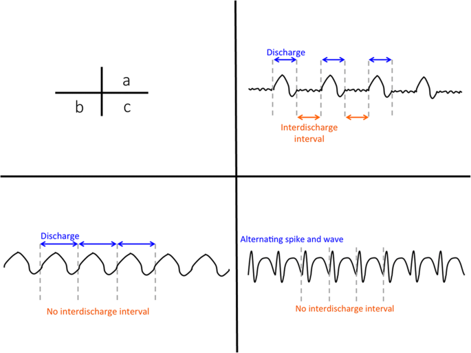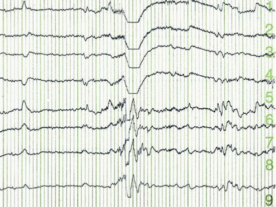

Through an EEG, doctors can look for abnormal patterns that indicate seizures and other problems. Normal electrical activity in the brain makes a recognizable pattern. Small metal discs with thin wires (electrodes) are placed on the scalp, and then send signals to a computer to record the results. If you have partial seizures, spikes and sharp waves on the EEG in a specific area of the brain. Übersetzungen für EEG seizure patterns im Englisch-Deutsch-Wörterbuch, mit echten Sprachaufnahmen, Illustrationen, Beugungsformen. An EEG tracks and records brain wave patterns. Though uncommon, they can occur in healthy persons without a history of seizures. Certain other patterns indicate a tendency toward seizures. The presence of IED in a routine EEG in children with a new-onset seizure is 18% to 56%, while in adults, it is 12% to 50%. Electrographic seizures: Although there are exceptions, the primary features which identify an EEG pattern as a (definite) seizure are: distinct beginning and end evolution in frequency and morphology or any regular rhythmic or repetitive pattern at >3Hz. They have a low sensitivity in routine 30 minute EEG recording, and the yield increases with repeat EEG and prolonged EEG recordings. EEG pattern are more likely to portend poor prognosis than seizures seen during rewarming.

They represent the epileptic focus in patients with seizures. Unfortunately, there remain many EEG patterns of indeterminate. Interictal epileptiform discharge is an abnormal synchronous electrical discharge generated by a group of neurons in the region of the epileptic focus. They are seen in patients with altered levels of consciousness. It is hypothesized that they occur due to structural or metabolic abnormalities at the thalamocortical levels due to the changes in the thalamocortical relays. Triphasic waves are seen diffusely with bifrontal predominance and are synchronous. The first phase is always negative, hence the name triphasic waves. They are sharply contoured with three phases. They are high amplitude sharp waves, with the duration of each phase longer than the next. However, these are nonspecific and can be seen in any metabolic encephalopathy. Triphasic waves were first believed to be pathognomic of hepatic encephalopathy. Triphasic waves: Triphasic waves were initially described in 1950 by Foley, and in 1955 Bickford and Butt gave it the name. The proposed approach employs Empirical mode decomposition (EMD) in order to decompose non-stationary EEG signals into intrinsic mode functions (IMFs). Medications like sedatives (phenobarbital, benzodiazepines) commonly cause diffuse beta activity. This paper introduces a new discriminant feature-Multi-level local patterns (MLP) for classification of seizure and seizure-free electroencephalogram (EEG) signals. Focal beta activity sometimes seen in structural lesions and also in various epilepsies (generalized fast activity/GFA). Beta activity is present in the frontal regions of the brain and can spread posteriorly in early sleep. Delta waves can be seen in drowsiness and also in very young children however, the appearance of focal delta activity can be abnormal (see below). However, in certain comatose states, there can be diffuse alpha activity (alpha comma) and may be considered pathognomonic.

For example, alpha waves are seen over the posterior head regions in a normal awake person and considered as the posterior background rhythm. The electroencephalographer is expected to have the significant skills to recognize artifacts, and also an understanding of normal, benign variants. This article reviews the abnormal waveforms in EEG recordings.Įven normal EEG waveforms can be considered potentially abnormal, depending upon various factors. Deep electrical activity of the brain is not well sampled in an EEG using extracranial electrode monitoring.Ībnormal waveforms seen in an EEG recording include epileptiform and non-epileptiform abnormalities. In order to identify abnormal waveforms in EEG, the reader should have a basic understanding of the normal EEG pattern in various physiological states in children and adults. It represents fluctuating dendritic potentials from superficial cortical layers, which are recorded in an organized array pattern and require voltage amplification to be captured. It is a tracing of voltage fluctuations versus time recorded from multiple electrodes placed over the scalp in a specific pattern to sample different cortical regions. Decision Points are designed to guide you through key health decisions, combining medical information with your personal information to make a wise health decision.Electroencephalography (EEG) was first used in humans by Hans Berger in 1924.


 0 kommentar(er)
